
Bronchial Wall Thickening: Causes and Is It Dangerous?
Bronchial wall thickening is a finding on a chest X-ray or CT scan, not a disease. Learn what causes it, how it is treated, and whether it is dangerous.
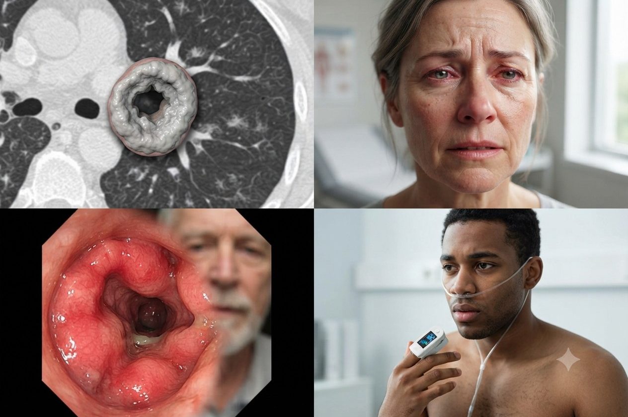
On this page
Bronchial wall thickening is not something you can see or feel yourself. It is a sign that shows up only on imaging tests, such as a chest X-ray or a CT scan. Conditions linked to this sign include airway inflammation, infections, pulmonary fibrosis (scarring of the lungs), asthma, and some inherited diseases.
So what is bronchial wall thickening, and is it dangerous? This guide explains the causes, how doctors look for the underlying problem, and how it is treated.
What is bronchial wall thickening?
The bronchi are the main airways of the lungs. Their walls are lined with a layer of mucus that helps keep foreign particles and germs from moving down into the lungs. In a healthy person, the bronchial walls cannot be seen on a chest X-ray. When too much mucus builds up in the bronchial walls, they can show up clearly and look abnormally thick on both sides of the lungs on an X-ray or CT scan.
Thickened bronchial walls on a scan can be a sign of many conditions. The most common is inflammation of the airways. You cannot see or feel this change yourself. Only a doctor can identify it, by looking at imaging tests.
What causes bronchial wall thickening?
The most common causes are inflammation, conditions a person is born with (for example, cystic fibrosis and alpha-1 antitrypsin deficiency), and obstructive lung diseases, which narrow or block the airways.
Asthma
Asthma is a chronic (long-term) disease of the airways. Inflammation and tightening of the muscles around the airways narrow them. Less air flows through, and the person has to work harder to breathe.
Signs of asthma include:
- Shortness of breath and trouble breathing
- Chest pain or tightness
- Wheezing
- Waking up at night with trouble breathing or a dry cough
Some people have worse symptoms when they have a cold or the flu, or when the weather changes. Other triggers can include smoke and dust, grass, pollen, animal fur, harsh soaps, and perfumes.
Asthma does not have specific features on a chest X-ray. If a person has no other lung disease, the X-ray is almost always normal. When changes do appear, they are bronchial wall thickening and overinflated lungs. A CT scan can show thickened bronchial walls along with narrowed airway openings and air trapped in the lungs when breathing out.
Bronchitis
Bronchitis is an infection of the main airways of the lungs (the bronchi) that leaves them irritated and inflamed. Causes include bacteria, viruses, cigarette smoke, air pollution, and other irritants.
The most common symptom is a cough. It can be dry, or it can bring up thick yellow-gray phlegm, although this does not always happen. Other symptoms include:
- Headache
- Fever
- Dry cough or cough with phlegm
- Sore throat
- Runny or stuffy nose
- Body aches
- Fatigue
Bronchitis can be acute (short-term) or chronic (long-term). Chronic bronchitis is a form of chronic obstructive pulmonary disease (COPD). Its symptoms include wheezing, shortness of breath that gets worse with exercise or movement, a lasting cough with phlegm, and frequent infections.
A chest X-ray in acute bronchitis usually shows nothing unusual. It is often ordered to tell bronchitis apart from pneumonia. Sometimes the X-ray shows thickened bronchial walls.
Cystic fibrosis
Cystic fibrosis is an inherited condition in which a faulty protein affects the cells, tissues, and glands that make the body’s mucus and sweat. Normal mucus is slippery and helps protect the airways, the digestive tract, and other organs and tissues. In cystic fibrosis, mucus becomes thick and sticky. When it builds up, it can cause blockages, damage, or infections in the affected organs.
The lungs are the organs most often affected. Some people with cystic fibrosis wheeze and cough up mucus, which may contain blood. Other symptoms depend on which organs are affected. The main signs of later-stage cystic fibrosis are bronchiectasis and thickened bronchial walls.
Bronchiectasis
Bronchiectasis is a chronic condition in which the airways of the lungs become widened. Extra mucus then builds up, which makes the lungs more likely to get infections. It develops after the lungs have been damaged by inflammation or infection. Symptoms include:
- A long-lasting cough with thick, foul-smelling phlegm
- Coughing up blood
- Shortness of breath and wheezing
A chest X-ray is often not enough to diagnose and guide treatment of bronchiectasis. The image may look normal, or it may show widened bronchi with thick walls. A high-resolution CT scan is the “gold standard” for diagnosis. It gives a clearer picture of the widened airways and the thickened bronchial walls.
Alpha-1 antitrypsin deficiency
Alpha-1 antitrypsin deficiency is a condition that raises the risk of lung disease and other diseases. Alpha-1 antitrypsin is a protein made in the liver that helps protect the lungs. If your body does not make enough of it, your lungs are more easily damaged by smoking, pollution, or dust in the environment. This can lead to COPD, bronchiectasis, or another lung disease.
The symptoms are similar to those of asthma and COPD. Some people have no symptoms at all.
Other causes
- Lung disease related to smoking or to work exposures
- Fungal lung disease
- Obstructive sleep apnea
- Cancer and hyperplasia (excess growth of cells)
How is bronchial wall thickening diagnosed and treated?
Once thickened bronchial walls are found on imaging, your doctor may order other tests to look for the cause. There is no specific treatment for bronchial wall thickening itself. Instead, the underlying cause is treated.
- Asthma: Quick-relief medicines and, if needed, controller medicines. Allergy treatment if the asthma is triggered by allergies. Surgery may be an option for severe asthma that does not improve with medicine.
- Bronchitis: Medicines to relieve symptoms if a virus is the cause. Antibiotics are added if bacteria are the cause.
- Bronchiectasis: Treatment mainly focuses on keeping the airways clear. Antibiotics are used during flare-ups, and exercise is also part of treatment.
- COPD: Bronchodilators (medicines that open the airways), inhaled steroids, antibiotics, oral corticosteroids, regular vaccinations, pulmonary rehabilitation, oxygen therapy, and surgery for severe cases.
- Alpha-1 antitrypsin deficiency: There is no specific treatment yet, but there are ways to slow lung damage. If a person has emphysema and the protein deficiency, standard COPD treatments can be used.
Ask your doctor which treatment is right for you.
Is bronchial wall thickening on both sides dangerous?
In most cases, this finding is linked to inflammation and infection of the airways. Once those conditions are controlled, it is usually not dangerous. Sometimes, however, thickened bronchial walls can be linked to chronic conditions that put your overall health at risk.
If you notice unusual signs in your body, it is best to see a doctor for an exam and treatment.
References
- Bronchial wall thickening — Radiopaedia
- Chronic Respiratory Symptoms Associated With Airway Wall Thickening Measured by Thin-Slice Low-Dose CT — American Journal of Roentgenology
- Asthma — Radiopaedia
- Cystic fibrosis (pulmonary manifestations) — Radiopaedia
- What Is Cystic Fibrosis? — NHLBI (NIH)
- Bronchiectasis — Radiopaedia
- Alpha-1 Antitrypsin Deficiency — NHLBI (NIH)
Medical disclaimer
FAQHealth content is for informational and educational purposes only. It is not a substitute for professional medical advice, diagnosis, or treatment. Always talk with your doctor or another qualified health professional about any questions you have about a medical condition.